We bring here a novel way to analyze and evaluate asthmatic reaction of histological images with computer vision techniques.
Abstract
We bring here a novel way to analyze and evaluate asthmatic reaction of histological images with computer vision techniques. A system named BEARI was developed throughout this project in order to help medical researchers to improve histological images analysis. This project is a part of an interdisciplinary medical research, cooperated with HADASA Medical School in Jerusalem.
The problem
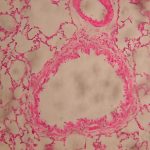
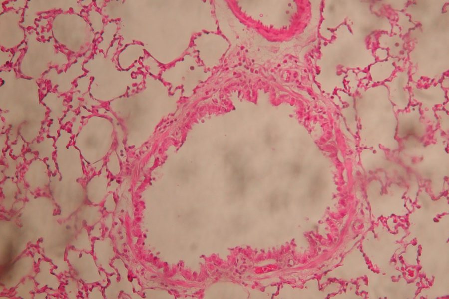
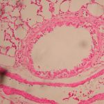
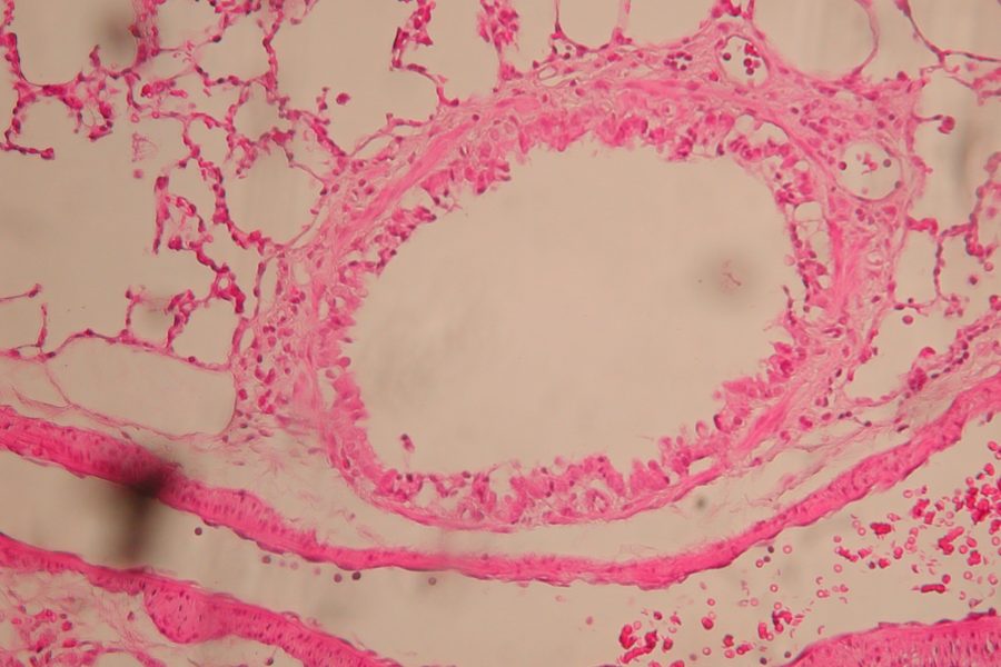
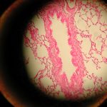
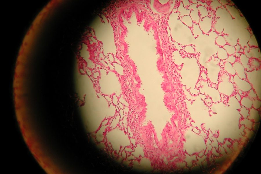
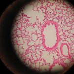
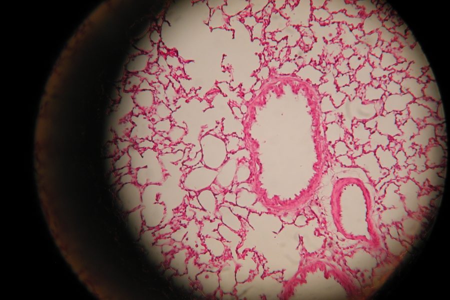
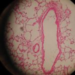
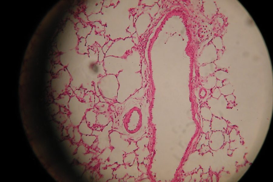
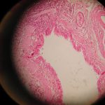
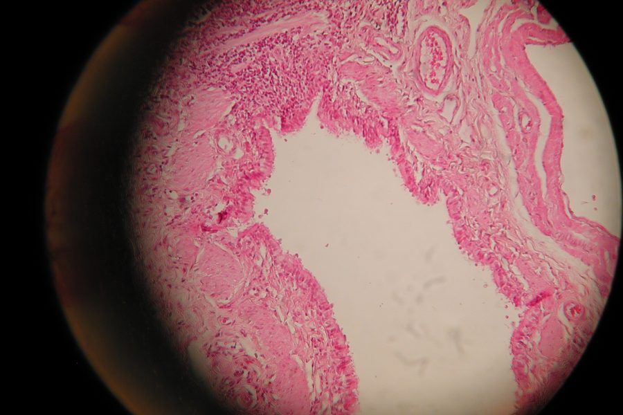
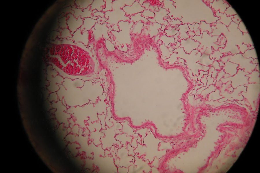
Asthma is one of the common epidemics of the respiratory system in human beings. One way to explore the influence of the asthmatic reaction on the lung tissue is using histological images. Analyzing those pictures is a complex mission for human eyes due to numerous cells examination and multi parameters consideration. This action is being held only by specialists, and it consumes valuable time and produces inconsistency in the results. In addition, results are measured into strict rank, and are lack of fine division. Use of computerized system will provide consistency, objectivity and will shorten the analysis time. Effective tool would even help in further researches.
The solution
The solution comes in a form of a computerized tool based on two image processing approaches. The Morphological approach, appointed for the definition of the Region Of Interest (ROI) in the image, and the Chromatic approach appointed for the segmentation of the inflammatory cells.
The BEARI system includes four modules as seen in the following figure ROI Recognition

ROI Recognition
The first module finds the Region Of Interest, which is the area that consists the asthmatic epidemic cells. This module takes raw pictures and performs image enhancement. After bronchus selection and validity check, the inner and outer ROI is being automatic recognized.
Parametric Analysis
The second module role is to find all important data for the asthmatic evaluation in the ROI. The module takes the recognized ROI and performs another image enhancement. Then the following operations are being held: Chromatic clustering, objects and background separation, Segmentation and statistical segments filtering. At its output we get the cells marked image & cells statistics.
Manual Selection
Manual selection Module is a stand alone tool that enables the user to manually mark the cells in the image. This tool was developed to enable next module evaluation.
Comparison & Statistics
The last module compares between the auto detection that was done by the Parametric analysis and the manual marking by the user. Together with the Manual Selection this module serves as a reliability test and quality check of the systems.
The system is designed with a friendly and easy to use GUI combines all system abilities, hence the name:
BEARI – Graphical User Interface

Tools
The images were taken by Nikon CoolPix 990 (3.2MP) under magnification of x20 with regular light microscope.
The computer vision system was implanted using Matlab 7.1 service pack 3 and its image processing toolpack.
Conclusions
BEARI is a powerful tool which demonstrates the benefits of computer vision techniques and how they can aid medical research. The results show how fast and consistent computer analysis is compared to human analysis. Analyzing histological images can be used in many different medical applications and areas, such as real time analysis of biopsy during tumor removal operations.
Acknowledgment
We are grateful to our project supervisor Eli Appelboim for his help and guidance throughout this work. We’re also grateful for the devoted work of the medical stuff: our project partner on the medical side Dr. Shai Paz, and the professional guidance of Dr. David Shusaiov. We also thank the Ollendorff Minerva Center for its support.